Leg Bones Diagram : Leg Bone Diagram. Learn how to draw the femur, patella, tibia, and fibula in this lesson! It is sometimes called the lower leg. The patella in the knee; However, the definition in human anatomy refers only to the section of the lower limb extending from the knee to the ankle, also known as the crus or. The femur, or thighbone, is the longest and largest bone in the human body.
Human anatomy diagrams show internal organs, cells, systems, conditions, symptoms and sickness information and/or tips for healthy living. The patella in the knee; Learn vocabulary, terms and more with flashcards, games and other study tools. It mainly serves as an attachment point for the muscles of the lower leg. The humerus and the femur are corresponding bones of the arms and legs, respectively.
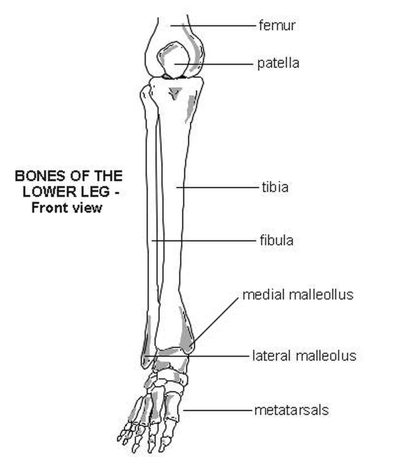
1000+ images about Anatomy and Physiology on Pinterest | Muscle atrophy, Human hand bones and ... from s-media-cache-ak0.pinimg.com Other joints, such as those between the vertebrae in. The tarsal bones in the ankle; The femur, or thighbone, is the longest and largest bone in the human body. Health diagram bone skeleton leg knee science anchor chart human human body. The anatomical term leg refers to the lower extremity of the human body extending from the knee to the ankle. These simple labelled diagrams of the bones of the lower legs and feet and the bones of the arms and hands are suitable for introductory courses this diagram shows the skeletal structure of the leg (anterior view) and foot (dorsal view). Blood vessels and nerves enter the bone through the nutrient foramen. Learn vocabulary, terms and more with flashcards, games and other study tools.
File is ready to render.
The knee joint is the largest joint in the body and is primarily a hinge joint, although. Human anatomy diagrams show internal organs, cells, systems, conditions, symptoms and sickness information and/or tips for healthy living. This diagram depicts diagram leg bones anatomy. Most bones (particularly the long bones of the arms and legs — which make up the appendicular skeleton) have a hard outer shell known as cortical bone. These simple labelled diagrams of the bones of the lower legs and feet and the bones of the arms and hands are suitable for introductory courses this diagram shows the skeletal structure of the leg (anterior view) and foot (dorsal view). At the microscopic level, this hard outer shell is made up of rod like structures called osteons. Ankle and foot pain massage therapy connections. Learn vocabulary, terms and more with flashcards, games and other study tools. While some people with paget's disease have no symptoms, others figure 9. Time to jump right into the biggest and strongest bones in the human body. All of your bones, except for one (the hyoid bone in your neck), form a joint with another bone. 12 photos of the diagram of leg bones. It is sometimes called the lower leg.
Diagram of blood and nerve supply to bone. Click on the figures for a detailed view and nomenclature. High quality realistic skeleton legs. The femur in the thigh; All of your bones, except for one (the hyoid bone in your neck), form a joint with another bone.
Human Skeleton Labeled Diagram . Human Skeleton Labeled Diagram Parts Of The Arm Bone Diagram ... from i.pinimg.com File is ready to render. This diagram shows the bones of the femur and the patella. Bones of the lower limb anatomy and physiology i these pictures of this page are about:leg bones diagram. 12 photos of the diagram of leg bones. Visit kenhub for more skeletal system quizzes. The knee joint is the largest joint in the body and is primarily a hinge joint, although some sliding and rotation occur. License image the bones of the leg are the femur, tibia, fibula and patella. Learn how to draw the femur, patella, tibia, and fibula in this lesson!
He leg's main function in the human is for locomotion and support of the rest of the body.
Here are a few anatomical plates about the leg and the foot. Learn vocabulary, terms and more with flashcards, games and other study tools. Health diagram bone skeleton leg knee science anchor chart human human body. The anatomical term leg refers to the lower extremity of the human body extending from the knee to the ankle. Quizzes on human skeletal system anatomy, bone anatomy, and bone markings. He leg's main function in the human is for locomotion and support of the rest of the body. At the same time, the bones and joints of the leg and foot must be strong enough to support the body's weight while remaining flexible enough for movement and balance. Want to learn more about it? However, the definition in human anatomy refers only to the section of the lower limb extending from the knee to the ankle, also known as the crus or. You will find the pelvic bones in the hip; This diagram depicts diagram leg bones anatomy. Joints hold your bones together and allow your rigid skeleton the bones in your skull are held together with fibrous connective tissue. Includes leg (femur, tibia, patella, and fibula) and foot (tarsals and digits) bones.
While their parts are similar in general, their structure has been adapted to differing functions. The foot bones shown in this diagram are the talus, navicular, cuneiform, cuboid, metatarsals and calcaneus. This diagram shows the bones of the femur and the patella. The humerus and the femur are corresponding bones of the arms and legs, respectively. Learn vocabulary, terms and more with flashcards, games and other study tools.
Pictures Of Bones Of The Lower Extremities from healthiack.com Learn vocabulary, terms and more with flashcards, games and other study tools. Learn how to draw the femur, patella, tibia, and fibula in this lesson! Its lower end helps create the knee joint. Visit kenhub for more skeletal system quizzes. The foot bones shown in this diagram are the talus, navicular, cuneiform, cuboid, metatarsals and calcaneus. All of your bones, except for one (the hyoid bone in your neck), form a joint with another bone. Here are a few anatomical plates about the leg and the foot. Ankle and foot pain massage therapy connections.
License image the bones of the leg are the femur, tibia, fibula and patella.
The human leg consists of 8 bones, 4 per leg. The knee joint is the largest joint in the body and is primarily a hinge joint, although some sliding and rotation occur. Other joints, such as those between the vertebrae in. It is sometimes called the lower leg. The bones of the leg are the femur, tibia, fibula and patella. At the same time, the bones and joints of the leg and foot must be strong enough to support the body's weight while remaining flexible enough for movement and balance. Want to learn more about it? He leg's main function in the human is for locomotion and support of the rest of the body. Joints hold your bones together and allow your rigid skeleton the bones in your skull are held together with fibrous connective tissue. While their parts are similar in general, their structure has been adapted to differing functions. Also, they provide an environment for bone marrow , where the blood cells are created, and they act as a storage area for minerals, particularly calcium. Learn how to draw the femur, patella, tibia, and fibula in this lesson! Time to jump right into the biggest and strongest bones in the human body.
Share :
Post a Comment
for "Leg Bones Diagram : Leg Bone Diagram"


Post a Comment for "Leg Bones Diagram : Leg Bone Diagram"